

![]()
Geographic distribution of the specimens
The genus Aotus shows a widespread geographic distribution from Panamá and the northern part of South America to South Paraguay, extending through the Amazonian tropical forest. In Peru, A. nancymai is found mostly South of the Amazonas-Solimões-Marañón river, while A. vociferans is encountered north of the same river system (Hershkovitz, 1983).
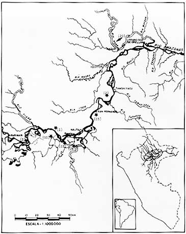 |
Figure 1 - Map showing the collecting sites of the specimens investigated, black stars corresponding to those where Aotus nancymai and arrow to that where Aotus vociferans were capture. Numbers in parenthisis indicate sample sizes. No information about the collecting site of 15 A. nancymai specimens from the east bank is available. |
Twenty specimens of A. vociferans were captured on the west bank of the Amazon river (Napo river) and 93 specimens of A. nancymai at several locations along the two banks of the Marañón-Amazon river (see Figure 1). Specimens were identified by the taxonomic criteria proposed by Hershkovitz (1983) to distinguish Aotus species (Aquino and Encarnacion, 1988). The animals were trapped by personnel of the Peruvian Primatological Project "Manuel Moro Sommo" and kept at the Centro de Reproducción y Conservación de Primates (CRCP) in Iquitos, Peru.
Sample collection
All specimens were anesthetized with an intramuscular injection of ketamine hydrochloride (10 mg/kg of body weight). Salivation was induced by an intramuscular injection of 0.001 ml of pilocarpin chlorhydrate (Hertape). After collection, saliva was immediately heated to 100ºC, on a water bath for 20 minutes to prevent glycoprotein hydrolysis. Five ml of blood were collected by puncture of the femoral vein using EDTA as an anticoagulant. Blood and saliva were centrifuged, and the supernatants stored on ice and shipped to the Belém laboratory.
Serological tests
Saliva samples were tested for the presence of A, B and H substances by the hemagglutination inhibition test using human anti-A and anti-B antibodies from Ortho Corporation, both at 1:256 titers, and Ulex europeus lectin (anti-H) prepared in our laboratory at 1:64 titer and both read after incubation at room temperature (see Schneider et al. , 1987 for a detailed description of procedures). Anti-A and antiB antibodies were diluted to about 8 agglutinating units for human A2 cells and B cells, while anti-H was adjusted to 4 units for O human cells. Cells from the same donor were used in all tests. Monkey sera were tested against human Al, A2, B and O cells by the hemagglutination test after absorption with human O red cells to remove non-specific antibodies.
Statistical analysis
All gene frequencies were estimated by the maximum likelihood method using the MAXLIK program prepared by Reed and Schull (1968). Fisher's test or the G test were used to verify differences of phenotypic frequencies between the two species. Differences between means were examined by the Student t-test using the SPSS program (Statistical Package for Social Sciences).
