
Short Papers
J. Med. Primatol. 19:151-153 (1990)
![]()
Trichobezoars in Two Saddleback Tamarins (Saguinus fuscicollis)
![]()
Alfonso S. Gozalo,1) Enrique Montoya, 1) Thomas E. Nolan 2)
1)Peruvian Primatological Project, Iquitos, Peru; 2)Merck Sharp & Dohme Research
Laboratory, West Point, PA, USA
Two captive-born Saddleback tamarins (Saguinus fuscicollis) died unexpectedly in the primate colony at the Peruvian Primatological Project. At necropsy, a firm, mobile, oblong, obscure mass was discovered in the stomach of each. They were removed and determined to be trichobezoars.
Key words: Aberrant appetite *Pathology e Callitrichidae
INTRODUCTION
![]()
There is scarce literature reporting cases of trichobezoars in nonhuman primates. Phytobezoars have been reported in three Langur monkeys (Pygathrix nemaeus) [3], causing intestinal obstruction and perforation prior to death. Two rhesus monkeys (Macaca mulatta) have been reported as having gastric phytobezoars [2] and one was reported to have a ligonobezoar [1]. Recently, there was a report of a gastric trichobezoar in a chimpanzee during routine surgery [4].
CASE HISTORIES
Two Saguinus fuscicollis monkeys captive born at the Center for Reproduction and Conservation of Nonhuman Primates at Iquitos, Peru, were housed as a breeding pair in an open-system breeding unit. The open system consisted of concrete- and wooden-frame wire-covered pens contained within a block construction screened in a building. Pens were approximately four feet square by six feet high. The animals had been maintained on a standard in-house prepared, grain-based, baked diet, fruit supplementation, and water ad libitum.
© 1990 Munksgaard
Case One
A 30 month-old male animal, identified as F-30, was found dead in its cage. At necropsy, the animal was found to be in generally good condition with the exception of having a sparse and short hair coat. Further exploration revealed a firm, oblong, noncompressible mass measuring three centimeters in width and five centimeters in length located in the lumen of the stomach. The mass consisted of matted hair.
Case Two
A 21 month-old female identified as F-37 presented with weight loss, sparse and short hair coat, lethargy, and anorexia. The animal died a few hours after being placed in an intensive-care incubator. At necropsy, a similar mass as described in case one was found in the stomach and extending into duodenum. The mass measured three centimeters in width and six centimeters in length. It consisted of matted hair (Fig. 1).
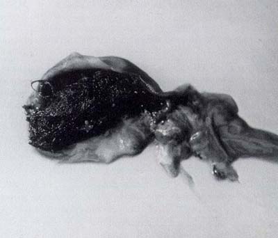
Fig. 1. Saguinus fuscicollis stomach showing a gastric trichobezoar
